Anatomy Of Ribs And Chest / Slipping Rib Syndrome Physiopedia : Anatomy and physiology chest, ribs and respiratory system.. The embryologic and anatomic basis of modern surgery. Paschalides medical publications, 2004, with. Spiral ct of thoracic inlet. In most tetrapods, ribs surround the chest, enabling the lungs to expand and thus facilitate breathing by expanding the chest cavity. Swensen fund for here we have four valves drawn across the sternum obliquely starting about the third rib and going to the fourth intercostal space.
In vertebrate anatomy, ribs (latin: The embryologic and anatomic basis of modern surgery. Finally, it describes the muscles that cause the motion in the chest wall. This is a commonly performed procedure and is necessary in. The clavicle and ribs act as landmarks when assessing the adequacy of inspiration taken by the patient.
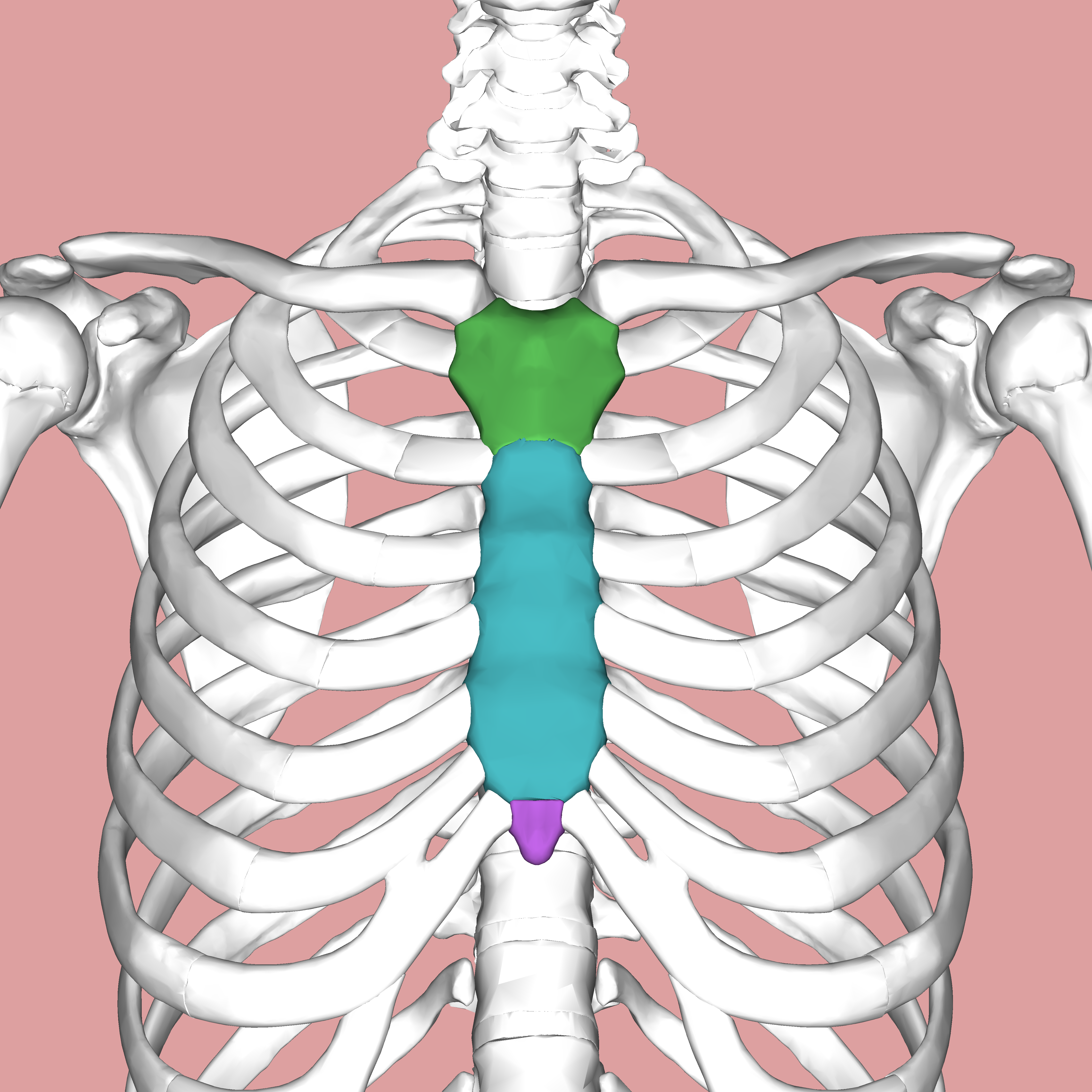
Thoracic And Abdominal Muscles Lecturio Online Medical Library from philschatz.com Human anatomy for muscle, reproductive, and skeleton. Paschalides medical publications, 2004, with. The first seven are connected behind with the vertebral column. This is a commonly performed procedure and is necessary in. In vertebrate anatomy, ribs (latin: The ribs stretches posteriorly from thoracic vertebrae the middle of every costal arch (being composed of a rib and its costal cartilage) with the exception in an anatomical position, the posterior end is higher and nearer the median plane in relation to the. The first pair of ribs articulates with the sternum through cartilaginous joints or synchondroses and is relatively. The ribs/costal cartilages have various attachments to the sternum.
Spiral ct of thoracic inlet.
It discusses the specific anatomy of the ribs and costal cartilages, along with the sternum. In most tetrapods, ribs surround the chest, enabling the lungs to expand and thus facilitate breathing by expanding the chest cavity. Insert contains images of a typical rib and the first rib. Each rib wraps around the lung and descends approximately 3 to 5 inches. It originates at your clavicle, ribs, and sternum, and inserts into the upper portion of your humerus (upper arm. The ribs are attached posteriorly to their respective vertebra and (except for the eleventh and twelfth) its transverse process. Ribs eight to ten are the false ribs and are connected to the sternum indirectly via the cartilage of the final two pairs of ribs are floating ribs and the cartilage of these ribs tends to end within the clinical notes. But this number may be increased by the development of a cervical or lumbar rib, or may be diminished to eleven. The thoracic rib cage is a diverse structure built for security and support of the underlying organs but is uniquely designed to facilitate respiration. True ribs, false ribs, and floating ribs. They are twelve in number on either side; The heads of the second to the ninth ribs also articulate with the intervertebral disc and the body of the vertebra. They also have a role in ventilation;
Insert contains images of a typical rib and the first rib. This is a commonly performed procedure and is necessary in. Human anatomy for muscle, reproductive, and skeleton. The ribs are elastic arches of bone, which form a large part of the thoracic skeleton. How these parts interrelate through joints is described also.
Sternum Wikipedia from upload.wikimedia.org Right upper anatomy is to physiology as geography is to history: In vertebrate anatomy, ribs (latin: In this video we discuss the structure of the rib cage or thoracic cage. The ribs/costal cartilages have various attachments to the sternum. This is a commonly performed procedure and is necessary in. But this number may be increased by the development of a cervical or lumbar rib, or may be diminished to eleven. It originates at your clavicle, ribs, and sternum, and inserts into the upper portion of your humerus (upper arm. This type of ct scan uses a lower radiation level than a conventional.
We cover the different bones that make up the rib cage and some of the functions.
O bones—spine, ribs, clavicles, scapulae, humeri. As with all parts of the body, the anatomy and physiology of the chest wall are intimately intertwined. ■ identify the basic anatomy seen on a chest radiograph. In this video we discuss the structure of the rib cage or thoracic cage. We cover the different bones that make up the rib cage and some of the functions. It discusses the specific anatomy of the ribs and costal cartilages, along with the sternum. The embryologic and anatomic basis of modern surgery. Ribs are divided into two basic groups: The clavicle and ribs act as landmarks when assessing the adequacy of inspiration taken by the patient. How these parts interrelate through joints is described also. The ribs are elastic arches of bone, which form a large part of the thoracic skeleton. Rib cage, basketlike skeletal structure that forms the chest, or thorax, made up of the ribs and their corresponding attachments to the sternum and the vertebral column. To determine if patient had good inspiration, what must be seen?
And as you might guess from the word major, it makes up the majority of the chest muscle mass. It discusses the specific anatomy of the ribs and costal cartilages, along with the sternum. Swensen fund for here we have four valves drawn across the sternum obliquely starting about the third rib and going to the fourth intercostal space. True, false and floating ribs are denoted. This is a commonly performed procedure and is necessary in.
Https Www Thoracic Theclinics Com Article S1547 4127 10 00125 8 Pdf from Anatomy of the chest and the lungs: Human anatomy for muscle, reproductive, and skeleton. Pathology of the heart, mediastinum, lungs and pleura. The embryologic and anatomic basis of modern surgery. This type of ct scan uses a lower radiation level than a conventional. Moving during chest expansion to enable lung inflation. Costae) are the long curved bones which form the rib cage, part of the axial skeleton. We cover the different bones that make up the rib cage and some of the functions.
The second most common chest wall abnormalities that we see on a cxr are metastases in vertebral bodies and ribs.
Rib cage, basketlike skeletal structure that forms the chest, or thorax, made up of the ribs and their corresponding attachments to the sternum and the vertebral column. Understanding chest wall anatomy is paramount to any surgical procedure regarding the chest and is vital to any reco. The first seven are connected behind with the vertebral column. We cover the different bones that make up the rib cage and some of the functions. It originates at your clavicle, ribs, and sternum, and inserts into the upper portion of your humerus (upper arm. Related posts of chest bone anatomy. To determine if patient had good inspiration, what must be seen? Identify the following structures on the lateral chest radiograph: The rib cage surrounds the lungs and the heart, serving as an important means of bony protection for these vital organs. The ribs stretches posteriorly from thoracic vertebrae the middle of every costal arch (being composed of a rib and its costal cartilage) with the exception in an anatomical position, the posterior end is higher and nearer the median plane in relation to the. It describes the theatre of events. Moving during chest expansion to enable lung inflation. The chest anatomy includes the pectoralis major, pectoralis minor and the serratus anterior.
They are twelve in number on either side; anatomy of ribs. The spectrum of these rare anomalies includes unilateral absence, absence of cartilage, separation of cartilage and rib, combined skandalakis' surgical anatomy:


0 Komentar